
Contrast-enhanced Ultrasonography of Renal Artery: Exploring the "Clues" of Hypertension
The patient, Mr. Wang, has hypertension for more than ten years and has been taking antihypertensive drugs for a long time. The hospital did routine ultrasound examination, suspicious Mr. Wang renal artery stenosis, followed by further contrast-enhanced ultrasound examination, a clear diagnosis of renal artery stenosis. So, how is Mr. Wang suffering from hypertension related to the kidney? Are there any other effects on the kidneys? What tests are needed to diagnose it?
I.The kidneys have a very close relationship with blood pressure:
Renal hypertension is the most common type of secondary hypertension, and the incidence of renal artery stenosis caused by renal atherosclerosis is high in the elderly, diabetes, coronary heart disease, hyperlipidemia and smoking patients. Renal artery stenosis can cause hypertension (new hypertension, or poor control of the original hypertension medication), proteinuria, decreased kidney function, and even renal failure. In the early stage, most patients' symptoms are not obvious, and at this time, if a clear diagnosis can be made, timely adjustment of individual treatment plan is very important for the development and prognosis of patients.
Second, how to detect renal artery stenosis early?
There are many diagnostic methods for renal artery stenosis, mainly divided into noninvasive examination and invasive examination. The noninvasive examination mainly includes ultrasound, CT angiography and magnetic resonance angiography. Invasive examination mainly refers to radial renal artery angiography, which is also the "gold standard" for the diagnosis of renal artery stenosis.
Color ultrasound examination is convenient and non-invasive, non-radioactive radiation, is often the preferred inspection method, and the further application of contrast-enhanced ultrasound, a new technology widely developed in recent years, makes the diagnosis of renal artery stenosis close to the "gold standard" level.
Renal artery contrast-enhanced ultrasound (CEUS) is used to inject ultrasound contrast agents (such as sonovir and sulfur hexafluoride microvesicles for injection) through the anterior elbow vein to perform enhanced real-time dynamic observation of renal artery throughout the process, which greatly improves the diagnostic accuracy of renal artery stenosis by ultrasound and the detection rate of congenital variations in renal artery. This kind of contrast agent has high safety, small dosage, only about 1ml/ kidney, no hepatorenal toxicity, and most of the metabolism can be excreted with respiration within ten minutes after injection. It is especially suitable for patients with renal insufficiency who are not suitable for iodine examination or who are allergic to iodine and cannot undergo renal artery CT angiography.
At present, renal artery contrast ultrasound is mainly used to examine hypertensive patients with suspected renal artery stenosis and elderly patients with renal insufficiency, which greatly improves the detection rate of renal artery stenosis, especially mild to moderate renal artery stenosis, and provides diagnosis and treatment basis for early clinical drug treatment or surgical intervention, thus helping to control blood pressure. Reduce the incidence of adverse cardiovascular events and end-stage renal disease.
Professor Guo Fajin, President of the Ultrasound Medicine Branch of the Chinese Geriatric Society, Professor Ren Junhong, Director General, Ma Na, Deputy Director General, led the team of the Ultrasound Medicine Department of Beijing Hospital (National Geriatric Center) in September 2017 to open the first renal artery contrast ultrasound clinic in China, working closely with a number of clinical departments, and the number of cases examined has reached more than 1,100. It has been recognized by clinicians and patients, and the technology has also been supported by a number of project funds, and won the attention and affirmation of peer experts. Recently, led by Professor Ren Junhong, the Chinese Expert Consensus on Methods and Procedures of Renal Artery Contrast Ultrasound (2021 edition) was released after many discussions and revisions by domestic well-known experts, and recommended opinions were put forward for the standardization of methods and procedures of renal artery contrast ultrasound.
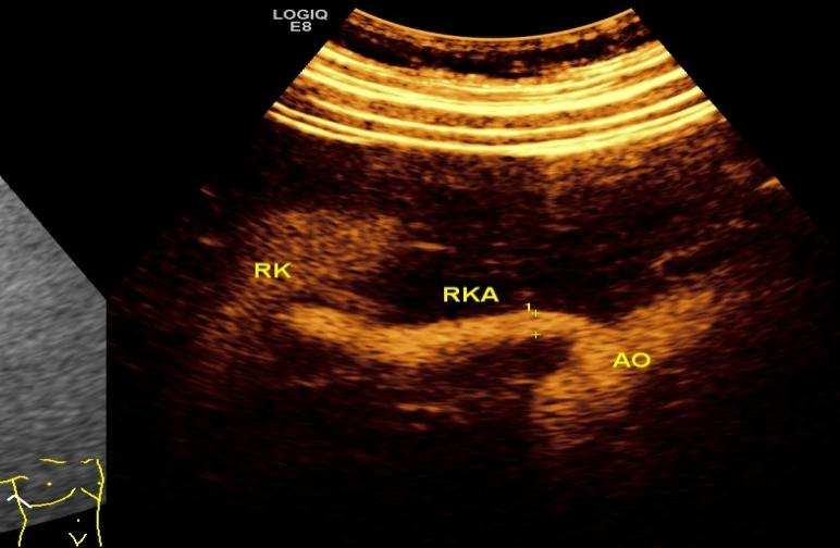 Figure 1 Contrast-enhanced ultrasound: Normal right renal artery throughout
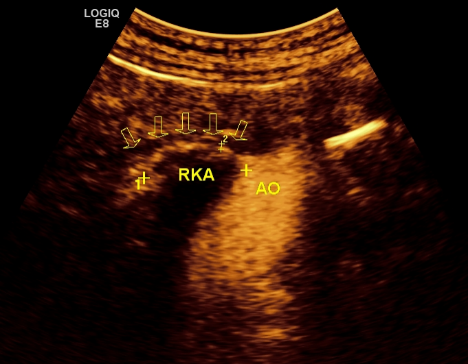 Figure 2. Contrasted ultrasound showing diffuse severe stenosis of the right renal artery
Author:
Department of Ultrasound Medicine, Beijing Hospital: Sun Youjing, Ma Na, Ren Junhong
(The opinions expressed are solely those of the author. Some pictures in this article are from the Internet, if there is infringement, please contact to delete)