
Enjoy the sunshine reasonably and be alert to precancerous lesions "solar keratosis"
The sea navigates by the helmsman, all things grow by the sun. Bathing in the sun, walking, dancing, playing chess, viewing, are very comfortable and relaxed; But enjoy the sun also need to have a degree, excessive long-term exposure, but may induce solar keratosis.
I.What is solar keratosis?
Solar keratosis (actinic keratosis, AK), also known as senile keratosis, is commonly seen in the elderly over the age of 65, is one of the most common skin diseases in the elderly group, is also a common preskin cancer, has the possibility of developing into invasive squamous cell cancer, so early detection and early treatment is crucial.
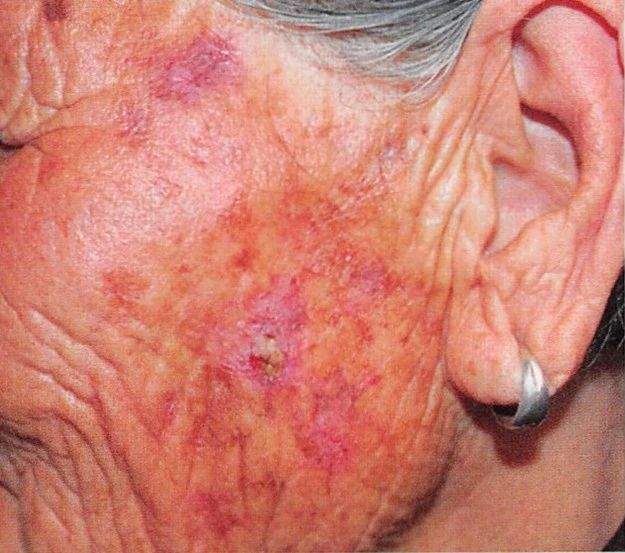
AKThe clinical manifestations are usually pale red or reddish brown papules or patches accompanied by scales, ranging from a few millimeters to two centimeters in diameter, often fused into sheets with a sandpaper-like texture to the touch. [1] The lesions were single or multiple. Multiple lesions were more common. Most are asymptomatic, with occasional mild pain and itching. According to the thickness of the lesion and the degree of keratosis, it is divided into 3 grades (Table 1) [2].
Table 1 Olsen clinical grade of photolinear keratosis
| fractionation
|
clinical picture.
|
| Ilevel
|
Light, light red to gray spots, a few scales, can be easily touched, not easily observed
|
| IIlevel
|
Moderate, erythema with more pronounced scales that can be easily reached and observed
|
| IIIlevel
|
Severe, thick squamous, pronounced hyperkeratosis, marked keratosis is easily observed, and early cutaneous squamous carcinoma is difficult to distinguish
|
| Regional carcinogenesis
|
Multiple AK lesions fused into slices
|
Two.What are the pathogenic factors that induce solar keratosis?
The pathogenic factors of AK include environment and individual factors. Environmental factors mainly refer to ultraviolet exposure, which can cause cell gene mutation, chronic skin inflammation, immune suppression, and eventually lead to abnormal proliferation of keratinocytes. The more UV the skin absorbs, the higher the risk of AK. Individual risk factors were age, sex, skin type, history of skin tumor and outdoor work history. People with fair skin (Fitzpatrick type I and type II skin) are more likely to develop AK. Immunosuppressed people, such as those with organ transplantation or long-term use of cytotoxic drugs, are more likely to develop AK and progress to cSCC[2].

Three.How to detect solar keratosis early?
If abnormal erythema or plaque is found in the exposed area, it is recommended to seek medical attention as soon as possible, and to confirm the diagnosis by dermatoscopy and skin biopsy under the guidance of a physician.
Among them, the sensitivity and specificity of dermatoscopy in the diagnosis of AK are high, which are 98.7% and 95%, respectively [3]. The three typical features under dermoscopy are red pseudoreticular structure, hair follicle horn plug, and strawberry sign [4]. Because of its intuitive, portable, non-invasive; High specificity and sensitivity; It can be used for differential diagnosis, monitoring treatment endpoint and follow-up observation [5].
Histopathological examination is still the gold standard for the diagnosis of AK, especially when the diagnosis is not clinically confirmed or invasive cSCC is suspected. The main indications for pathological examination were: unknown clinical diagnosis, lesions > 1 cm in diameter, bleeding, ulceration or induration, rapid growth of skin lesions; Secondary signs: Lesions accompanied by intense itching, pain, and obvious hyperkeratosis. Some special parts of the skin (such as lips) need to undergo pathological examination due to the high risk of invasion. In addition, pathological classification is helpful to guide the treatment and prognosis of AK [2].
Four.How to treat solar keratosis?
Solar Angle into precancerous lesions, has not yet developed into cancer, early detection, active cooperation with doctors treatment, the general prognosis is good. Follow-up monitoring is required after treatment.
The existing treatment methods mainly include local treatment and systemic treatment. Local treatment includes photodynamic therapy, local topical drug therapy, physical therapy (CO2 laser, cryotherapy), and local surgical excision. Physical therapy and local surgical excision are only used to treat existing lesions. Photodynamic therapy, local topical drug therapy can directly remove skin lesions, and can also be used for regional treatment.
Systematic treatment is mainly based on oral retinoic acid drugs, which is suitable for the regionalized treatment of multiple AK regional cancer or high-risk patients [2].


Five, how to prevent solar keratosis?

Because solar keratosis may become cancerous, attention should be paid to sun protection and excessive exposure to ultraviolet rays. High-risk patients, such as multiple skin lesions, old age, immunosuppression, previous tumor history and special skin lesions, need lifelong follow-up and pay attention to regional treatment. It is suggested that on the basis of clinical observation and follow-up, auxiliary diagnostic tools such as dermoscopy should be used for early detection and early standardized treatment, which is crucial for preventing the disease and preventing its progression.
References:
[1] He Li, Wu Xiaoqing, Li Jiangbin, et al. Research progress of solar keratosis [J]. Chinese Journal of Leprosy and Dermatology Journal of Science, 2021, 37(1): 60-64.
[2] Dermatology Rehabilitation Committee of Chinese Rehabilitation Medical Association, Photodynamic Therapy Research Center of Dermatology and Venereology Branch of Chinese Medical Association, Photomedical Treatment Equipment Group of Dermatology and Cosmetology Branch of Chinese Medical Equipment Association. Expert consensus on the clinical diagnosis and treatment of photolinear keratosis in China (2021)[J]. Chinese Journal of Dermatology Journal of Science, 2021, 54(12): 1048-1056.
[3] HUERTA-BROGERAS M, OLMOS O, BORBUJO J, et al. Validation of dermoscopy as a real-time noninvasive diagnostic imaging technique for actinic keratosis[J]. Arch Dermatol, 2012, 148(10): 1159-1164.
[4] ZALAUDEK I, GIACOMEL J, ARGENZIANO G, et al. Dermoscopy of facial nonpigmented actinic keratosis[J]. Br J Dermatol, 2006, 155(5): 951-956.
[5] Gao Yao-ying, Yang Jing, TAO Juan. Application value of skin imaging technology in the diagnosis and treatment of solar keratosis [J]. Chinese Medical Journal ,57, 2022 (9) : 947-949.
(The opinions expressed are solely those of the author. Some pictures in this article are from the Internet, if there is infringement, please contact to delete. Patient pictures shall not be used without authorization of the author and the platform.)
Author:
Gong Jin
Hubei Jingzhou First People's Hospital Dermatology
Young member of Dermatology Branch of Chinese Geriatric Society