
Does ophthalmological check-up check eyesight namely? NO!
";Teacher, my eyesight is very good, threading needles have no problem, what is the need to check the eye!"; "
";My vision is 5.2 in both eyes, it is a pilot's vision, and those slit lamp examinations and fundus photos are completely unnecessary!";
- Routine eye examination items include E word eye chart examination, slit-lamp microscope examination, fundus examination and intraocular pressure examination, but many medical customers think that eye examination is to check a vision so simple, so give up or ignore the routine eye examination, and this is likely to lose the opportunity to find some eye diseases "clues", a fat thing? Let me tell you more
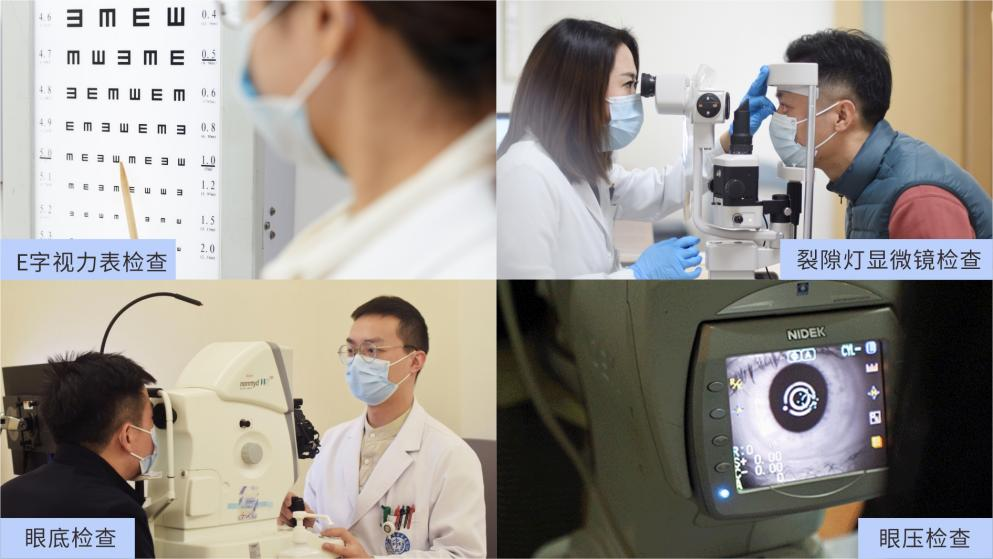
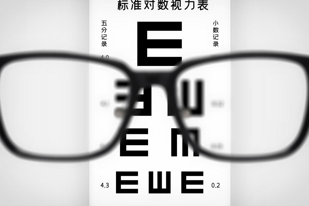
First, check the eye chart
The visual acuity chart is mainly to check the central visual acuity, which reflects the visual sensitivity of macular fovea, and has very important clinical significance for the screening of eye diseases. At present, there are two kinds of E-word visual acuity charts in use in China, namely, international standard visual acuity chart and standard logarithmic visual acuity chart. The standard logarithmic visual acuity chart is more scientific, and it is also the commonly used visual acuity chart in domestic eye medical institutions.
So, older brothers and sisters, whether you are a unit medical examination or personal medical examination, as long as there is an eye medical examination this project, you must carefully test vision, wear glasses elder brother some also need to wear glasses to test and correct vision oh.
2. Slit lamp microscopy
Slit lamp examination, also known as slit lamp microscopy, is to illuminate the examination site with a concentrated light source, and then cooperate with binocular micromagnifying glasses to form a series of "optical sections" through the transparent tissue of each part of the eye, so that different levels of the diopter stroma and even the small lesions of deep tissue can be clearly displayed. From front to back examination includes ocular skin, eyelashes, eyelids, conjunctiva, cornea, sclera, anterior chamber, aqueous humor, iris, pupil, lens and other intraocular tissues. The diagnostic examination of eye diseases such as conjunctivitis, keratitis, stes, and cataracts that we are usually concerned about is the credit of the slit lamp.
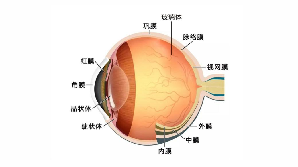
Figure 3: Eye structure
Third, fundus examination

Expression package1
In fact, this is not the case, because most of the current physical examination institutions use non-mydriatic fundus photography, so compared with the traditional direct ophthalmoscopy under small pupil, non-mydriatic fundus photography can more quickly and visually observe the fundus, especially the images of the posterior retina. Fundus photography is an important method to detect diseases of vitreous, retina, choroid and optic nerve, such as bleeding, exudation, retinal degeneration, retinal hiatal hole, retinal neovascularization, atrophic plaques and pigment disorders.

Expression package2
First of all, even if you are a pilot vision, it is better to regular Routine screening under the fundus, after all, your car is in regular maintenance, you say, the window of your mind should also take a look at it regularly?
Secondly, studies have shown that 6.7% to 32% of diabetic patients have varying degrees of diabetic retinopathy at the time of diagnosis. Diabetic retinopathy is the main cause of new forms of blindness in adults aged 20 to 65 years. Therefore, the primary screening of diabetic retinopathy is the first step to intervene in diabetic blindness. Patients with diabetes, especially those with a course of disease of more than 5 years, it is best to conduct a fundus examination every six months, and pull the first line of defense of sugar web screening.
In addition, patients with hypertension, hyperlipidemia, cardiovascular and cerebrovascular diseases are also high-risk groups of fundus diseases, such groups of people are recommended to check the fundus at least once a year, early screening, early detection, early treatment.
Moreover, for patients with cataract, glaucoma, high myopia and other eye diseases, regular follow-up should be carried out according to the doctor's advice.
Finally, for people with healthy eyes, people over 40 years old should check the fundus once a year, and people under 40 years old should check the fundus once every 1-2 years.
Iv. Intraocular pressure examination
Now that we're clear about the fundus examination, let's talk about the intraocular pressure examination. Intraocular pressure examination is to check the pressure inside the eyeball. Normal people's intraocular pressure fluctuates between 10 and 21mmHg. If the intraocular pressure is higher than 21mmHg, it is called intraocular hypertension, which is more common in optic nerve damage or glaucoma, and needs early and timely treatment.
Having said all this, you may have to ask:

Expression package3
If you suffer from conjunctivitis, stele, dry eyes and other ocular surface diseases for years, if you want to find out whether you have cataracts, or even the position of intraocular lenses after cataract surgery, and whether there are some complications, you at least have to do a slit lamp examination, if you also happen to suffer from diabetes, hypertension, hyperlipidemia or cardiovascular and cerebrovascular diseases, then, I suggest you take it seriously and get all your eye exams done.
Finally, tap on the blackboard:

Expression package4
I hope everyone can take good care of their "soul window" oh.
References:
1. Lin KY, Hsih WH, Lin YB, Wen CY, Chang TJ. Update in the epidemiology, risk factors, screening, and treatment of diabetic retinopathy. J Diabetes Investig. 2021 Aug; 12 (8) : 1322-1325.
2. Liu G. Basis of ophthalmology. 3rd edition [M]. Beijing: People's Medical Publishing House [in Chinese], 2010:252-257.
3. Yang Peizeng, Fan Xianqun. Ophthalmology. 9th Ed. [M]. Beijing: People's Medical Publishing House [in Chinese], 2018:286-290.
Writing:
Liu Zhenrong, attending physician
Health Management Center, West China Hospital, Sichuan University
Review:
Huang Yan, deputy chief physician
Vice President, Health Management Branch, Chinese Geriatric Society
Health Management Center, West China Hospital, Sichuan University